NEXT LEVEL coaching company begins to introduce you to how our brain works. First, let's talk about what the human brain is made of.
The human brain is a complex network of neurons. These neurons serve to build the nervous system. It is with their help that information is transmitted from the brain to the entire body and back.
You probably think that such a complex process requires a huge number of neurons. But how many neurons are there actually in the human brain? Previously, scientists, as one claimed: “100 billion,” but recent studies have shown that there are much fewer neurons in our brain.
How many neurons are there in the human brain?
According to many scientists, the human brain consists of about 100 billion neurons (give or take a couple of billion). It is this figure that has been cited for many years in textbooks on neurobiology and psychology. And all this time, scientists believed that this figure was close to the truth.
That was until Brazilian researcher Suzanne Herculano-Housel discovered that this figure was not entirely accurate. The scientist realized that despite the fact that data on 100 billion neurons are persistently published in many works, it is simply impossible to calculate where this magic figure came from. Then the neuroscientist decided to conduct her own investigation to finally find out how many neurons there actually are in the human brain.
It would seem that the task is elementary. Simply take a sample of the brain, count the number of neurons in that sample, and then purely mathematically calculate the total number of neurons, given the total volume of the brain.
What is a neuron (neural connections)
Translated from Greek, neuron, or as it is also called neuron, means “fiber”, “nerve”. A neuron is a specific structure in our body that is responsible for the transmission of any information within it, in everyday life called a nerve cell.
Neurons operate using electrical signals and help the brain process incoming information to further coordinate the body's actions.
These cells are an integral part of the human nervous system, the purpose of which is to collect all signals coming from outside or from one’s own body and make a decision about the need for one or another action. It is the neurons that help cope with this task.
Each of the neurons has a connection with a huge number of the same cells, a kind of “web” is created, which is called a neural network. Through this connection, electrical and chemical impulses are transmitted in the body, bringing the entire nervous system to a state of rest or, conversely, excitation.
For example, a person encountered a certain significant event. An electrochemical impulse (impulse) of neurons occurs, leading to the excitation of the uneven system. A person’s heart begins to beat faster, their hands sweat, or other physiological reactions occur.
We are born with a given number of neurons, but the connections between them have not yet been formed. The neural network is built gradually as a result of impulses coming from outside. New impulses form new neural pathways; it is along them that similar information will flow throughout life. The brain perceives each person's individual experience and reacts to it. For example, a child grabbed a hot iron and pulled his hand away. This is how he developed a new neural connection.
A child develops a stable neural network by the age of two. Surprisingly, from this age those cells that are not used begin to weaken. But this does not in any way interfere with the development of intelligence. On the contrary, the child learns about the world through already established neural connections, and does not aimlessly analyze everything around him.
Even such a child has practical experience that allows him to cut off unnecessary actions and strive for useful ones. This is why, for example, it is so difficult to wean a child from the breast - he has formed a strong neural connection between the attachment to mother's milk and pleasure, safety, and calmness.
Learning new experiences throughout life leads to the death of unnecessary neural connections and the formation of new and useful ones. This process optimizes the brain in the most efficient way for us. For example, people living in hot countries learn to live in a certain climate, but northerners need a completely different experience to survive.
Does the brain remember everything? Conversation with neurophysiologist Olga Svarnik
Image title
Today we have access to a wide variety of scientific instruments and the most advanced technologies. Humanity has accumulated colossal knowledge, both in the natural sciences and in the humanities. However, the human brain remains the “Holy Grail” of scientists and the most complex, little-studied area. What prevents us from studying the brain to the end? How does human memory work and does our brain really remember everything? Olga Evgenievna Svarnik , a neurophysiologist, candidate of psychological sciences, senior researcher at the Laboratory of Psychophysiology named after V.B. Shvyrkova Institute of Psychology RAS.
We all know that the brain is a very complex structure. Tens of billions of neurons, trillions of synapses... Given this complexity, how much are we able to study the brain and what is the main stumbling block in such research today?
We can certainly study the brain. And this is a rather long process, due to the features that you are talking about: a huge number of cells, connections, the cells are all very different. Research over the past decades has shown that there are a huge number of types of neurons, and the deeper we dive into this field, the more new types we find. The process of researching the brain and the cells that make up this brain is almost endless and very interesting.
An important question is how is the brain connected to mental processes? The activity of our neurons is related to what the body is doing. What's remarkable is not only that there are many different types of neurons in the brain, but also that they fire at specific moments that are specific to those neurons. There are neurons that will be active when I talk to someone about the brain, or when I myself think about how the brain works, or even when I sleep and dream about something about the brain. By examining these neurons, we gain access to the inner world of a person.
The main stumbling block in studying the brain is that the huge amount of detail that we obtain about the work of the brain for some reason does not want to fit into some generally accepted theory. And there are some changes in what we understand by how the brain works. There are several different proposals for what these brain principles are. And quite a large number of researchers cannot come to a consensus on this issue. There are a lot of details, but the overall picture has not yet emerged . We can see a similar situation in other sciences, for example, in physics.
Olga Evgenievna, you study memory. Tell us more about this. Is memory located somewhere in the brain or is it a situational process and we do not have a specific memory area?
In short, yes, there is no memory zone. At the same time, the destruction or disruption of certain areas can lead to amnesia. But it's not the same thing. There is short-term memory, there is long-term memory, there is implicit memory, when we have acquired some experience, but we cannot say anything about it and cannot somehow declare it. And there are types of memory where we can say, for example, that we know where the Eiffel Tower is or imagine how neurons work in the brain. These are all different aspects of the phenomenon that is commonly called memory. And when we talk about these manifestations of the brain, we cannot say that memory lies somewhere in a certain place in the brain.
One famous amnesiac named Henry Molaison had surgery to destroy the hippocampal structures and some of the cortical areas that were connected to the hippocampus, leaving him unable to remember anything. He did not have the impression that he could describe any episodes that happened to him. But at the same time, his training still took place, he just could not declare the episodes. Roughly speaking, the patient had information about the episodes, but he simply could not talk about it. And this phenomenon was described 50 years before the case of Henry Molaison. The Swiss doctor Eduard Claparède described a very famous, almost anecdotal case. He constantly shook hands with a patient of his with a similar disorder. The woman also had problems acquiring new memory and the ability to declare episodes from her life. During one of these greetings, the doctor placed a needle in his arm and injected the patient. Subsequently, the patient did not remember this at all, but began to avoid shaking hands with the doctor. It turns out that this experience still remained with the person, and such experience could shape the further interactions of this woman with the world.
In 2021, Olga Svarnik published the popular science book “The Brain in a Minute.”
Is it possible to say that our brain does not forget anything at all, and that what happened once remains forever?
The tendency in modern neuroscience is that the problem of memory is primarily a problem of access to it. The point is not that the memory was somehow lost. If we imagine that any acquired experience is the formation of some kind of neural group that is now associated with it, then it turns out that returning to this experience means activating this group. If we layer more and more other neural groups, moving away in our experience from that original group, then it turns out that we cannot return to it due to the fact that there are already other layers there and the branches of this “tree of experience” have grown considerably.
Experiments on animals show that it is possible to activate the old group that existed before all these layers and return to that moment. And in this sense, of course, we can say that yes, the brain really stores everything if there was an established experience . Around us now there are a lot of short-term moments, which for some short period are also “fixed” by our brain, but at the same time do not move into the long-term phase. But if everything has already passed into long-term memory, then the possibility of losing such a memory is, first of all, the difficulty of finding access to it, or another option is if the cells associated with this memory are destroyed.
How to explain the cases when some smell takes you back to such distant times that you seem to no longer remember, but suddenly the memory comes to life again? Is smell a realm of the subconscious? And how is it related to memory?
Most of what is in our brain works without reaching the level that is commonly called consciousness. But it still constitutes our experience.
In terms of the ability to return to the old neural groups of the experience that was before all the layers, smell plays a universal and very interesting role. That is, the smell helps to revive what we ourselves can no longer get close to: due to the connection of previous experience with many other things that we have become acquainted with in the course of life.
Why is this happening? To be honest, I don’t know the answer to this question, but it has interested me for a long time. Even a picture extremely rarely leads to such a revival of episodes from our past, but smell has such a unique opportunity. This phenomenon has been described many times and colorfully in fiction, but from a scientific point of view it is difficult to imagine what it could be. Why does the smell, not even the sound, have such characteristics? I would also like to know the answer to this question.
Why can we remember for many years some minor details from the distant past that seem to carry no meaning (for example, green socks seen on someone a long time ago, or a dog running past)? Or, as Freud said, there cannot be insignificant details and behind this memory there is some more serious, hidden experience?
Such memories are associated with some general state of the body at that time. Perhaps that state, in its emotional characteristics, really had great significance. Probably, this feature of our memory played a role in evolution: organisms that recorded as many details as possible with the help of their neurons probably received a greater advantage in evolution.
Another aspect is that the state, so important at that moment, could return again and again when we mentally think about the experience. And at the time of one of these returns, these green socks or something else could have been added. Perhaps they were not directly related to that situation, but our memory, after some time, connecting it and layering something else, “decided” that these green socks were very important for that situation. There are various nuances regarding how our memory undergoes various modifications with each reactivation of the neural groups that underlie it. These are also very interesting processes.
It turns out that, in fact, the most accurate memory is the first one, and all other returns, memories of this moment that were layered later, are false? Maybe all our memories are generally incorrect and have little connection with what actually happened?
It is very important to say what these types of memory are. The phenomenon of memory remodeling due to a return to the activation of the earliest neural group is still associated with episodic memory. But semantic memory works the other way around: if we are learning something, for example, trying to remember all the capitals of the world, then repetition is only beneficial and it strengthens our memory. (Semantic memory refers to knowledge (for example, that the Eiffel Tower is in Paris), and not the episode itself of my first vision of the Eiffel Tower). But the episode itself, which we witnessed, tends to change, acquire details that were not there, and lose those that were. Numerous studies suggest that episodic memory may not be given much credit. Whether the episodes from our childhood were exactly as we remembered them is not so simple a question. It may well be that there were similar things, but they looked completely different from how we remembered them.
Lecture by Olga Svarnik “Sleep and Memory” at the Bench RAS.
Olga Evgenievna, you work at the Institute of Psychology of the Russian Academy of Sciences, at the Moscow Institute of Psychoanalysis, and are actively teaching. What attracts you most as a scientist to neuroscience?
As a teacher, I talk about the principles of the brain to various students: from physicists to psychologists. As a scientist, I study the cells that are present in the brain at a variety of levels: these are neurogenetic changes, and changes in electrical activity, as well as changes in overall brain activity recorded using an electroencephalogram in humans.
I am very fascinated by the description and study of behavior, as well as the search for some common patterns between people and animals. My research shows that the process of acquiring some kind of experience (when the body is faced with some new situation for it) leads to the fact that we, first of all, reactivate those neural groups that are associated with something similar: already existing previous experience.
An interesting pattern is observed - how often and consistently we, when acquiring something new, return to the old. Both people and animals, having acquired new experience and found a solution to a new situation, having already added something new to their brain, return again and again to the old: to previously acquired forms of behavior. It’s as if they are testing the old behavior model over and over again, trying to make sure that it really doesn’t work? Didn't you work before? Experiments show that people often do not even realize this. And the question of how far we are returning to the old and why we are doing this is what occupies me most now.
Video title
Interviewed by Yanina Khuzhina.
How many neurons are in the brain
Nerve cells make up about 10 percent of the brain, the remaining 90 percent are astrocytes and glial cells, but their task is only to serve neurons.
Calculating the number of cells in the brain “manually” is as difficult as finding out the number of stars in the sky.
Nevertheless, scientists have come up with several ways to determine the number of neurons in a person.:
- The number of nerve cells in a small part of the brain is calculated, and then the number is multiplied in proportion to the total volume. Researchers start from the postulate that neurons are evenly distributed in our brain.
- All brain cells dissolve. The result is a liquid in which cell nuclei can be seen. They can be counted. In this case, the service cells that we mentioned above are not taken into account.
As a result of the described experiments, it was established that the number of neurons in the human brain is 85 billion units. Previously, for many centuries, it was believed that there were more nerve cells, about 100 billion.
The number of neural connections in the brain improves a person's quality of life
For many years, scientists thought that the adult brain remained unchanged. However, science now knows for sure: throughout our lives, more and more synapses are formed in our brain - contacts between neurons or other types of cells that receive their signals. In total
neurons and synapses form a neural network, the individual elements of which are constantly in contact with each other and exchange information.
The neuron is responsible for memory
Researchers have uncovered how episodic memory and associations occur at the level of a single neuron and how...
02 July 13:40
It is neural connections that help different areas of the brain transmit data to each other, thereby ensuring vital processes for us: memory formation, production and understanding of speech, control of the movements of our own body. When neural connections are disrupted, which can happen as a result of diseases such as Alzheimer's disease or physical trauma, certain areas of the brain lose the ability to communicate with each other. As a result, it becomes impossible to perform any action, both mental (memorizing new information or planning one’s actions) and physical.
A team of researchers led by Stephen Smith from the Center for Functional Magnetic Resonance Imaging of the Brain at the University of Oxford decided to find out whether the total number of neural connections in the brain could somehow influence its overall functioning. During the study, scientists used data obtained as part of the Human Connectome Project
- a project launched in 2009. Its goal is to compile a kind of “map” of the brain, with the help of which it will be possible to understand which area of the brain is responsible for a particular process or disease, as well as how different areas of the brain interact with each other.
What was unique about the work of Stephen Smith's research group was that the scientists did not focus on connections between specific areas of the brain or on specific functions, but rather studied processes as a whole.
read more about the findings in the journal Nature Neuroscience
.
The study used the results of magnetic resonance imaging of 461 people. For each of them, a “map” was created that showed the total number of neural connections between all areas of the brain. In addition, each study participant filled out a questionnaire about their education, lifestyle, health, marital status and emotional state. In total, the questions touched on 280 aspects of human life.
The paralytic got up and walked
For the first time in history, a man whose both legs were paralyzed for five years regained the ability...
September 24 11:42
As a result of the work, it was possible to find out: the greater the number of neural connections present in the human brain, the more “positive” it is.
People whose brains were rich in connections between neurons tended to have higher education, had no problems with the law, strived to lead a healthy lifestyle, were in good psychological health and generally showed high levels of life satisfaction.
According to the authors of the study, the relationship between the number of neural connections and the quality of human life was so clear and strong that the scientists themselves were amazed by it.
The science department was able to contact the lead author of the work, Stephen Smith, and talk to him about the details of the work.
— Is it possible to give an accurate explanation of why the number of neural connections in the brain has a direct impact on a person’s quality of life: for example, to say that the number of connections somehow affects brain activity?
— No, it’s too early to talk about such cause-and-effect relationships, since all this is the subject of complex and multivariate correlation analysis. Therefore, we cannot yet say that a brain with many neural connections makes a person study several years longer (or vice versa - that many years of study increases the number of neural connections).
By the way, at the moment it is indeed possible to extend cause-and-effect relationships in both directions - this can be called a “vicious circle”.
- In that case, how are you going to break this “vicious circle”?
“The work we have done now—scanning the brain using magnetic resonance imaging—can only show how closely certain areas of the brain are connected to each other. It also reflects many other biological factors of lesser importance - for example, showing the exact number of neurons connecting these areas. But understanding how these connections affect a person’s behavior, mental abilities, and lifestyle is the main question facing the staff of the Human Connectome Project.
Why you should sleep on your side
To cleanse the brain of toxins accumulated during the day, you need to sleep on your side, scientists have found. Science Department...
16 August 12:40
— Stephen, is there a correlation between the number of neural connections in the brains of parents and children?
- But here I can answer unequivocally - yes. There is a lot of evidence that the number of neural connections, so to speak, is inherited. As part of our project, we are going to study this phenomenon in more depth. Although, of course, there are other important factors that affect the functioning of the brain and the formation of neural connections.
— Is it possible, at least theoretically, to somehow influence the number of neural connections and thus change the quality of a person’s life?
“It’s very difficult to talk about this in general terms. However, there are many examples where interventions in the functioning of the brain changed a person’s behavior or improved some individual indicators of his work. You can read about such an experiment, for example, in the journal Current Biology
: the article states that scientists, using micropolarization (a method that allows one to change the state of various parts of the central nervous system by the action of direct current. - Gazeta.Ru), managed to improve the mathematical abilities of subjects.
Another, simpler and more ordinary example can be given: we all know that training and practice in any type of activity help improve the performance of this very activity.
But learning, by definition, changes the neural connections of the brain, even if sometimes we are not able to detect it.
Regarding your question, the problem of global change in human behavior or abilities remains a large-scale and extremely interesting object of study.
Neuron structure
The figure shows the structure of a neuron. It consists of a main body and a core. Numerous fibers branch off from the cell body, which are called dendrites.
Powerful and long dendrites are called axons, which in reality are much longer than in the picture. Their length varies from a few millimeters to more than a meter.
Axons play a leading role in transmitting information between neurons and ensure the functioning of the entire nervous system.
The junction of a dendrite (axon) with another neuron is called a synapse. Dendrites, in the presence of stimuli, can grow so much that they begin to pick up impulses from other cells, which leads to the formation of new synaptic connections.
Synaptic connections play a significant role in the formation of human personality. Thus, a person with an established positive experience will look at life with love and hope, a person who has neural connections with a negative charge will become a pessimist over time.
The cytoarchitecture of the human brain is organized in such a way that more than 10 billion nerve cells, occupying a relatively small space and being formed into specialized structures, provide specific brain functions associated with the perception, processing and conduction of information, in accordance with which the body interacts with external environment based on high neuronal specificity and plasticity.
The basic structural unit of the nervous system is the neuron.
Different types of neurons are differentiated by the size and shape of the cell body, as well as by the length and degree of branching of its processes.
The cell body varies very widely in size - from 5 to 100 microns in diameter. It contains the following organelles: nucleus, mitochondria, endoplasmic reticulum (smooth and rough), located on the cisterns of the endoplasmic reticulum and in the free space of ribosomes and polysomes, the Golgi complex and various intracellular inclusions (glycogen granules, lipid droplets, accumulations of pigment particles in special neurons and etc.), vesicles, and lysosomes. Groups of parallel cisterns of the rough endoplasmic reticulum in the form of membrane-bound elongated cisterns with ribosomes attached to them form the Nissl substance (tigroid substance). The cytoplasm also contains neurofilaments and neurotubules (Fig. 3).
All of the listed ultrastructural organelles of the cell have certain functions. The nucleus is the substrate of the main genetic processes in the cell. Mitochondria provide energy metabolism - oxidative phosphorylation occurs in them, leading to the production of energy in the form of ATP molecules. The endoplasmic reticulum with ribosomes attached to its cisterns, as well as freely located ribosomes and their complexes (polysomes) are related to protein metabolism and synthetic processes in the cell. Lysosomes are assigned an metabolic and excretory role. Neurotubules and neurofilaments provide transport of intracellular substances related to the conduction of nerve impulses. For a long time it was believed that the Golgi complex, consisting of parallel cisterns and clusters of vesicles at their ends, performs undefined metabolic and excretory functions. Although not everything is known about this complex, the data accumulated by many researchers is attractive, indicating that it plays a major role in the processes of renewal of the cell membrane and its genetically determined specialization. It is known that in the Golgi complex the primary assembly of specialized sections of the membrane (receptors) can occur, which in the form of vesicles are transported to the outer cell membrane and are integrated into it. Such studies were summarized by A.A. Milokhin (1983).
The main process, the axon, and numerous branching processes, dendrites, extend from the body of the neuron. The length of the axons of various neurons ranges from 1 mm to almost 1 m (nerve fiber). Near the end, the axon divides into terminals on which synapses are located that contact the body and dendrites of other neurons. Synapses, together with neurofilaments and neurotubules, are the substrate for the conduction of nerve impulses.
Rice. 3. Basic ultrastructural components of a neuron.
L - lysosomes; RER - rough endoplasmic reticulum (cisterns with attached ribosomes); M - mitochondria; NF - neurofilaments; NT—neurotubes; P - ribosomes; P - polysomes (ribosome complexes); CG - Golgi complex; I am the core; CER - endoplasmic reticulum cisterns; LG - lipid granules; LF - lipofuscin.
In addition to neurons, brain tissue contains various types of glial cells - astroglia, oligodendroglia, microglia. Astroglia plays a major role in ensuring the function of the neuron and shaping the response of brain tissue to harmful influences (infection, intoxication, etc.) - it takes part in inflammatory processes and the elimination of their consequences (replacement gliosis). Oligodendroglia are known to provide myelination of nerve fibers and regulate water metabolism (drainage glia). The functions of microglia are not fully understood, but their importance is emphasized by the proliferation of these cells in some specific processes (participation in the formation of senile plaques; there is an assumption that microglial cells produce amyloid fibrils, etc.).
Special cellular structures are characteristic of the ventricular surfaces of the brain and its choroid plexus. The ventricular surface of the brain is covered with ependymal cells with numerous microvilli and cilia that take part in the circulation of the cerebrospinal fluid; The choroid plexus is represented by “clusters” of villi consisting of capillaries covered with epithelial cells. Their main function is related to the exchange of substances between blood and cerebrospinal fluid.
Typical synapse
consists of a presynaptic terminal, a postsynaptic region and a synaptic cleft located between them. The presynaptic terminal is the end of the axon. It contains neurofilaments, neurotubules, mitochondria and synaptic vesicles, clusters of which are visible near the presynaptic membrane. The neurotransmitters contained in the vesicles are transported through the latter. A postsynapse is characterized by the presence of a postsynaptic thickening. The postsynaptic thickening is represented by a cell membrane with receptors located on it, which are part of the structure of the membrane itself. The synapse is shown in Fig. 4, and its electron microscopic picture in Fig. 5.
A synapse can be located on the cell body (soma) - axosomatic synapse, on a dendrite - axodendritic, on a dendrite spine - axospinous (Fig. 6) and on the axon of another cell - axo-axonal. Axospinous synapses are somewhat different in structure from a typical synapse, which is determined by the structure of the spine, which has a special spiny apparatus as part of the postsynapse.
The interaction between the presynapse and postsynapse is ensured by the transfer of neurotransmitters across the synaptic cleft. Released from the presynapse, a neurotransmitter (mediator) can bind to a receptor on the postsynaptic membrane, be inactivated in the synaptic cleft, and be partially recaptured by the presynaptic membrane (reuptake process). If the receptor of the postsynaptic membrane is blocked, then both of the latter processes are possible, as well as excessive accumulation of the transmitter and the associated development of receptor hypersensitivity (see Fig. 4).
These processes are discussed in more detail in the section “Neurochemical systems of the brain.”
Receptors
neurons are protein structures located on the outer surface of the cell membrane.
They are able to “recognize” and bind biologically active substances - neurotransmitters, various endogenous substances, as well as exogenous compounds, including psychopharmacological agents. Compounds that can bind receptors are called ligands.
Ligands can be
endogenous
or
exogenous.
Ligand recognition by the receptor is ensured by special structural elements, or sites. The specificity of ligand binding occurs due to the structural correspondence of the ligand and receptor molecules when they fit together like a “key to a lock.” The binding reaction is the moment when a cascade of intracellular reactions is triggered, leading to a change in the functional state of the neuron. Depending on the “strength” and “strength” of binding of the ligand to the receptor, the concept of affinity
(affinity) of the ligand in relation to the receptor.
When a receptor binds to a ligand, both activation and blockade of the receptor can occur. In this regard, they talk about agonists
and
receptor antagonists,
as well as
partial agonists
(Fig. 7).
has maximum effectiveness in terms of receptor activation ,
minimal (almost zero) - antagonist.
In between are substances called partial agonists. The latter act much more mildly than full agonists. Partial agonists, in addition, occupying a certain spatial position in the receptor molecule, can prevent the excessive effect of the full agonist, i.e. act partially as antagonists. In this case, the concept of agonist/antagonist is used.
Both receptor agonists and antagonists can have high affinity. The agonist activates the receptor, causing the corresponding physiological effect, while the antagonist, by binding to the receptor, blocks it and prevents the development of the physiological effect detected by the agonists. An example of antagonists are antipsychotics, which prevent the effects of dopamine at the dopamine receptor level.
When a ligand binds to a receptor, the configuration of the latter changes (Fig. 7).
Many substances, both endogenous and exogenous, react not with one, but with several types of receptors - a “family” of them, which is divided into separate types. An example is the many neurotransmitters that react with several types of specific receptors (for example, D1-D5 types of dopamine receptors). The existence of several receptors for one ligand is called receptor heterogeneity.
An idea of the function of receptors would be incomplete if one did not imagine the intracellular processes that develop after the receptor binds to the corresponding substance, and the mechanisms that ensure the transformation of an external signal into processes leading to the appearance of a nerve impulse. Binding of a ligand to a receptor can lead either directly to the opening (or closing) of the corresponding ion channels (see Fig. 7) or to the activation of secondary messenger systems
(a substance that reacts with the receptor is considered as the primary messenger).
The first mention of secondary messenger systems appeared in connection with the work of E. Sutherland et al. (1950), who showed that adrenaline stimulates glycogenesis by increasing the concentration of cyclic adenosine monophosphate (cAMP) in the cell. It turned out that this second messenger mediates other cellular reactions. Subsequently, a connection was identified between the action of cAMP and the activation of protein kinases—enzymes that phosphorylate proteins, which leads to changes in their structure and activity.
Later, other secondary messengers were discovered. Now there are 3 classes among them: 1) cyclic nucleotides (cAMP, cyclic guanosine monophosphate - cGMP); 2) calcium ions (Ca2+); 3) phospholipid metabolites - inositol-1,4,5-triphosphate (1P3), diglycerol (DAG), arachidonic acid. Unlike other second messengers, Ca2+ is transported into the neuron from the intracellular space.
Neuron membranes contain specialized transmembrane proteins that form ion channels not only for Ca2+, but also for other ions, the concentration of which on both sides of the membrane affects changes in membrane potential. Polarization and depolarization of the membrane occur, i.e. change in transmembrane potential. The ion channels for sodium (Na+), potassium (K+), chlorine (C1-) and calcium (Ca2+) are of greatest importance in these processes.
Types of neurons and neural connections
Neurons can be found in various human organs, not exclusively in the brain. A large number of them are located in the receptors (eyes, ears, tongue, fingers - sensory organs). The collection of nerve cells that permeate our body forms the basis of the peripheral nervous system. Let us highlight the main types of neurons.
| Type of neuron cell | What is he responsible for? |
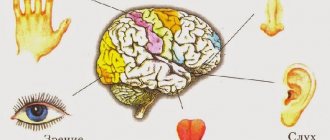
| Affective | They are carriers of information from the sense organs to the brain. This type of neuron has the longest axons. An impulse from the outside enters along axons strictly into a certain part of the brain, sound - into the auditory “compartment”, smell - into the “olfactory” compartment, etc. |
| Intermediate | Intermediate nerve cells process information received from affector neurons and transmit it to peripheral organs and muscles. |
| Effector | At the final stage, efferents come into play and carry the team of interneurons to the muscles and other organs of the body. |
The coordinated work of three types of neurons looks like this: a person “hears” the smell of kebab, the neuron transmits information to the corresponding section of the brain, the brain transmits a signal to the stomach, which secretes gastric juice, the person decides “I want to eat” and runs to buy kebab. This is simply how it works.
The most mysterious are the interneurons. On the one hand, their work determines the presence of a reflex: if you touch electricity, you pull your hand away, dust flies, you close your eyes. However, it is not yet clear how the exchange between fibers gives rise to ideas, images, thoughts?
The only thing that scientists have established is the fact that any type of mental activity (reading books, drawing, solving mathematical problems) is accompanied by a special activity (flare) of nerve cells in a certain area of the brain.
There is a special type of neurons called mirror neurons. Their peculiarity lies in the fact that they not only become excited by external signals, but also begin to “move”, observing the actions of their fellow neurons - other neurons.
Functions of neurons
Without neurons, the human body cannot function. We saw that these nanocells are responsible for literally our every movement, every action. The functions they perform have not yet been fully studied and defined.
There are several classifications of neuron functions. We will focus on what is generally accepted in the scientific world.
Information dissemination function
This function:
- is basic;
- studied better than others.
Its essence is that neurons process and transfer to the brain all impulses that come from the outside world or one’s own body. Next, they are processed, similar to how a search engine works in a browser.
Based on the results of scanning information from the outside, the brain, in the form of feedback, transmits the processed information to the senses or muscles.
We do not suspect that information is delivered and processed every second in our body, not only in the head and at the level of the peripheral nervous system.
To date, it has not been possible to create artificial intelligence that would come close to the operation of human neural networks. Each of the 85 billion neurons has at least 10 thousand experience-based connections, all of which work to transmit and process information.
Function of accumulating knowledge (saving experience)
A person has memory, the ability to understand the essence of things, phenomena and actions that he repeated once or many times. Neuronal cells, or more precisely neurotransmitters, connecting links between neighboring neurons, are responsible for the formation of memory.
Thus, it is not any separate part of the brain that is responsible for memory, but small protein bridges between cells. A person can lose memory when these neural connections collapse.
Integration function
This function allows individual lobes of the brain to interact with each other. As we have already said, signals from different sense organs arrive in different parts of the brain.
Neurons transmit and receive impulses in different parts of the brain through “bursts” of activity. This is how the process of thoughts, emotions and feelings appears. The more such diverse connections there are, the more effectively a person thinks. If a person is capable of reflection and analysis in a certain direction, then he will think well in another matter.
Protein production function
Neurons are such useful cells that they are not limited only to transmission functions. Nerve cells produce proteins necessary for human life. Again, neurotransmitters that are responsible for memory play a key role in the production of proteins.
In total, about 80 proteins are induced in neurons, here are the main ones that affect human well-being:
- Serotonin is a substance that causes joy and pleasure.
- Dopamine is the leading source of vigor and happiness for humans. Activates physical activity, helps to wake up, an excess can lead to a state of euphoria.
- Norepinephrine is a “bad” hormone that causes attacks of rage and anger. Along with cortisol, it is called the stress hormone.
- Glutamate is a substance responsible for memory storage.
Stopping the production of proteins or releasing them in insufficient quantities can lead to serious illnesses.
Basics of Brain Development
Over the past few decades, significant advances have been made in our understanding of the major stages and mechanisms of mammalian brain development. Research related to the neurobiology of brain development spans levels of brain organization from the macroanatomical to the cellular and molecular. This knowledge provides a picture of brain development as the product of a complex series of dynamic and adaptive processes operating within a constrained, genetically organized, but constantly changing context.
Human brain development is a long-term process that begins in the third gestational week (GW) with the differentiation of neural progenitor cells and extends through at least late adolescence, possibly throughout life. The processes that contribute to brain development range from molecular events of gene expression to environmental influences on the developing brain. These very different levels and types of processes interact to support the ongoing series of events that determine brain development. Both gene expression and environmental exposure to the brain are essential for normal brain development, and disruption of either can dramatically alter neurodevelopmental outcomes. But neither genes nor environmental influences prescribe or determine this outcome. Rather, brain development is precisely characterized as a complex series of dynamic and adaptive processes that operate throughout development to promote the emergence and differentiation of new neural structures and functions.
By the end of the embryonic period (end of the embryonic period - GW8), the rudimentary structures of the brain and central nervous system have already been defined and the main "compartments" of the central and peripheral nervous systems are formed. The subsequent period of fetal development continues until the end of pregnancy. During this time, rapid growth and development of both cortical and subcortical structures occurs, including the rudiments of the main fiber pathways (tracts). Changes in the overall morphology of the prenatal neural system are reinforced by changes occurring at the cellular level. Neuron production in humans begins at embryonic day 42 (E42), which is after 42 days from conception. In this case, neurons migrate to different areas of the brain, where they begin to communicate with each other, creating rudimentary neural networks. By the end of the prenatal period, the major fiber pathways, including the thalamocortical pathway, are completed.
Brain development continues over a long period of time. The brain quadruples in size during the preschool period, reaching approximately 90% of adult volume by age 6 years. But structural changes in the main compartments of gray and white matter (matter) continue in childhood and adolescence, and these changes in the structure of parallel changes and functional organization are reflected in the behavior of children and adolescents. During the early postnatal period, the level of neuronal connectivity throughout the developing brain far exceeds the level of neuronal connectivity in adults (Innocenti and Price 2005). This intense connection gradually weakens in its severity due to competitive processes that are influenced by the experience of the human body. Early experience-dependent processes underlie the plasticity and adaptability that are hallmarks of early brain development.
Before talking about brain development, it is worth touching on some concepts, in particular from the field of genetics. Genes are material substances that are passed on from generation to generation between generations. Genes are contained in nucleotide sequences of DNA (DNA), which are found in the nucleus of every cell in the body. Gene expression has one result: the production of a protein molecule. These molecular products of gene expression are essential for all aspects of development. Genes provide the template for the production of proteins, and it is proteins that are the active agents in biological development. Thus, although genes contain information that is essential for the development and functioning of a biological organism, genes are essentially inert molecules. Genes cannot directly participate in biological processes. Rather, there is an indirect relationship between the information in a gene and the developmental outcome. Information in gene sequences must be extracted, recoded, and translated into proteins. Proteins are part of complex interactive signaling cascades that typically involve multiple gene products as well as environmental influences. Thus, a particular gene product is one of many important elements that interact to support and guide the complex process of brain development.
Let's say a few words about the organization of the mature human brain. The human brain is perhaps the most complex of all biological systems. The mature brain consists of more than 100 billion neurons (Pakkenberg, Gundersen 1997). Neurons are essentially information processing cells in the brain. There are many different types of neurons, which differ in their size and shape, as well as their functions. Neurons connect with each other to form information processing networks that are responsible for all our thoughts, sensations, feelings and actions. Because each neuron can make connections with more than 1,000 other neurons, the adult brain is estimated by researchers to have more than 60 trillion neuronal connections. The point of communication between two neurons is called a synapse.
The mature human brain has a characteristic structure of sulci and convolutions. The spherical shape of the mature brain is believed to be an adaptation to the dramatic increase in brain size during evolution. The "folding" of brain tissue allowed relatively large brains to fit into relatively small cranial vaults, which had to remain small to accommodate the birth process. The brain's largest and most important information processing networks include the neocortex and subcortical nuclei, which relay information to and from the neocortex. The neocortex is a 2-5 mm thick layer of cells that lies on the surface of the brain (the word "cortex" comes from the Latin term meaning bark, as in the bark of a tree). In a cross-section of the brain, the neocortex is a thin, dark gray strip that follows the surface of the brain, like its mantle. The subcortical nuclei are clusters of neurons that serve as signaling relay centers communicating between the neocortex and the rest of the body, and as relays between different areas of the cortex. They are located deep in the brain below the cortex and are thus called "subcortical" nuclei. Because both the neocortex and subcortical nuclei contain the cell bodies of neurons, they have a gray appearance, giving rise to the term "gray matter." Populations of neurons are interconnected by fibers that extend from the cell bodies of individual neurons. There are two types of connective fibers: dendrites and axons. Dendrites are arrays of short fibers that resemble tree branches; collections of dendrites are often referred to in the literature as “dendritic arbors”. They extend only a short distance from the cell body of the neurons. Their main function is to receive electrochemical input signals from other neurons. Axons are long connecting fibers that extend over long distances and connect to other neurons, often at dendrites. Axons act like "telephone wires" because they are responsible for sending electrochemical signals to neurons located in distant locations. Bundles of individual axons from different neurons in one area of the brain form fiber pathways (tracts) that spread and connect to groups of neurons in other areas of the brain, forming information processing networks. The axons are "wrapped" in a fatty substance called myelin, which, like the insulation on a telephone wire, makes the transmission of electrochemical signals between areas more efficient. Myelin appears white in appearance, which is why the brain's fiber tracts are often referred to as "white matter" or "white matter tracts." In the very center of the brain there are a number of interconnected cavities that form the ventricular systems of the brain. The ventricular system is filled with a fluid called cerebral spinal fluid, which is completely recirculated several times a day. The ventricular system has a number of important functions, including cushioning and protecting the brain, removing waste, and transporting hormones and other substances (Brodal 2010). During brain development, the walls of the ventricles are the site of the greatest number of neurons. Although the neocortex of the brain may be relatively homogeneous in structure, it is actually divided into structurally and functionally distinct regions. These areas differ in the types of neurons they contain, the types of input they receive, and the types of connections they make to other brain regions. These structural differences lead to functional differences, creating areas of the brain that are specialized to perform different types of processes.
Let us now return to the topic of this article, that is, to the basics of brain development. In humans, the embryonic period begins at conception and spreads through GW8. By the end of the embryonic period, the rudimentary structures of the brain and central nervous system have already been determined and the main sections of the central and peripheral nervous system have been formed. The early embryonic period, which lasts until approximately mid-gestation, is a critical period in the development of the neocortex. By this time, most of the cortical neurons have been generated, and many of them have already migrated to their positions in the neocortex and began to process information using the main networks of the brain.
At the end of the second week after conception, the embryo is a simple, oval, two-layer structure. It is of interest to review the basic spatial dimensions of the embryo at "embryonic day" 13 (E13) and during the embryonic period of development it is often designated by the number of days after conception, which is called the embryonic day, so gastrulation begins with embryonic day 13 (E13). Each of the two layers contains a very primitive cell type. The top layer contains epiblast cells and the bottom layer contains hypoblast cells. By the end of the third week, the embryo has transformed through a series of processes collectively called gastrulation into a three-layer structure. Although it may seem like a simple change, the cell line transformations that occur during gastrulation set the stage for all subsequent stages of embryonic development. The epiblast cells of the upper cell layer will further differentiate into three primary stem cells, lineages that will ultimately give rise to all structures in the developing embryo, while the hypoblast cells of the lower layer will form extraembryonic tissues such as the fetal component of the placenta and the connective stalk. Among the stem cell lines that arise during gastrulation, neural stem cells are identified. Neural stem cells are capable of producing all the different cells that make up the brain and central nervous system, and for this reason neural stem cells are commonly called neural progenitor cells. The major gastrotation events occur between E13 and E20. The onset of gastrulation is marked by the formation of a primitive stripe and a primitive node. The initial streak provides the opening of the deeper embryonic layers. The first step in the gastrulation process is signaled by the appearance of a slit-like opening in the top layer of the embryo, called the primitive streak. It provides access to the lower regions of the embryo. A subset of epiblast cells then detaches from the top layer of the embryo and begins to migrate toward the primitive streak. When they reach the slit-like opening, they change direction and pass through the primitive streak and under the top layer. They then change direction again and begin to move towards the rostral end of the embryo. The rostral end of the embryo will later develop into the baby's head. The earliest migrating cells will move to the most rostral positions of the embryo, and then migrating cells will move sequentially to more caudal regions that will become the neck and trunk of the body. Migrating cells form two new embryonic layers. The cells that form the deepest layer will displace the hypoplast cells and form the endodermal stem cell layer, which will lead to the formation of intestinal and respiratory tract structures, while the cells that form the new intermediate layer of mesodermal stem cells will lead to the formation of such structures such as muscles, bones, cartilage and the vascular system. Cells that remain in the epidermal layer develop into one of two types of ectodermal stem cell layer. Epidermal ectodermal stem cells will lead to the formation of structures such as skin, nails and sweat glands, while neuroctodermal stem cells will lead to the development of the brain and central nervous system. Neuroectodermal stem cells are neural progenitor cells.
The differentiation of all embryonic stem cell lineages is associated with complex molecular signaling cascades. At the onset of gastrulation, the epiblast layer cells that will differentiate into neural progenitor cells are located along the rostral-caudal midline of the bilayer embryo. The differentiation of these cells into neural progenitor cells is the result of complex molecular signaling that involves several gene products (i.e., proteins) that are produced by several different populations of embryonic cells. Recall that at the beginning of gastrulation, epiblast cells begin to migrate in a certain direction and then pass through the primitive streak. As the subset of cells that migrate along the rostral-caudal midline of the embryo approach opening, they pass through another structure called the primitive ganglion, which is located at the rostral end of the primitive streak. The primitive node is a molecular signaling center. The cells of the primitive ganglion send a molecular signal to a subset of cells that migrate along the rostral-caudal midline of the embryo, and this signal in turn causes gene expression in the migrating cells. Expression of the gene in the migrating cell produces a protein that is secreted into the space between the migrating cells and the cells that remain in the midline region of the upper epiblast layer. The secreted protein binds to receptors on the surface of cells in the upper layer of the embryo and induces epiblast cells to differentiate into neural progenitor cells.
Thus, at the end of gastrulation, cells located along the midline of the upper layer of the embryo are transformed into neural progenitor cells. Differentiation of neural progenitor cells requires complex genetic signaling among at least three cell populations: node cells, migrating cells, and cells that will become neural progenitors. However, in reality this early signaling appears to be even more complex. In addition to producing molecular signals that induce migrating cells to produce proteins that are subsequently transformed into overlying epidermal cells, neural progenitor cells, the primitive ganglion generates another set of signals that changes during gastrulation and serves to form the basic rostral-caudal department of the embryonic nervous system. Recall that the earliest migrating epidermal cells move to the most rostral end of the embryo, and then the migrating cells move to larger caudal locations. The primitive ganglion sends signals to all migrating cells to produce proteins that signal neural progenitor cells, but each subsequent wave of migrating cells also receives a second signal indicating the regional identity for the neural progenitors. Thus, the primitive ganglion signals (initiates) early migration of epidermal cells to obtain molecular signals for cells in the overlying layer to differentiate into neural progenitors capable of producing cells suitable for forebrain structures, while later migrating cells signal differentiation neural progenitors capable of producing cells suitable for the hindbrain or spinal cord.
The next major step in brain development involves the formation of the first clearly defined neural structure, the neural tube, which forms in the third week of gestation, between E20-27. As noted earlier, by the end of gastrulation, neural progenitor cells have differentiated and are already located along the rostral-caudal midline of the upper layer of the three-layer embryo. The area of the embryo containing neural progenitor cells is called the neural plate. The first sign of neural tube development is the appearance of two ridges that form on either side of the neural plate around E21. Neural progenitor cells are located between the two ridges. Over the course of several days, the ridges rise, fold inward, and fuse to form hollow tubes (Copp et al., 2003). Fusion begins in the center of the developing neural tube and then occurs in both rostral and caudal directions. The anterior neuropores, at the most rostral end of the neural tube, and the posterior neuropores, at the caudal end, are the last segments to close at E25 and E27. When the neural tube is complete, neural progenitors form a single layer of cells that connects the center of the neural tube, immediately adjacent to its cavity by the centrum. In the embryo, the hollow center of the neural tube is cylindrical, like the center of a straw. But as the brain becomes larger and more complex, the shape of the cavity also changes, eventually forming the ventricular systems of the brain. Because neural progenitors are located in the region that will become the ventricles, this region is called the “ventricular zone” (VZ). Neural progenitor cells in the most rostral region of the neural tube will form the brain, while more caudally located cells will form the hindcord and spinal cord.
Although the basic three-dimensional organization of the embryo emerges with the formation of the neural tube, the embryo undergoes rapid growth over the next month. At the end of the neurulation period, the embryo is 3 to 5 mm long, and by the end of GW8 it increases to 27-31 mm, that is, ten times larger. During this period, the shape of the primitive nervous system changes dramatically. Just before the neural tube closes, the anterior end of the tube begins to expand, forming three primary brain vesicles (vesicles) or sacs. The largest portion of these embryonic brain vesicles is called the “prosencephalon,” which is the embryonic precursor of the forebrain. The middle vesicle is the "mesencephalon", which is the precursor of the midbrain structures, and the most posterior is the "rhombencephalon", which will later become the hindbrain. These three segments are further subdivided and by the end of the embryonic period five secondary brain vesicles are present. The prosencephalon is divided into the "teleencephalon" and the "diencephalon", and the rhombencephalon is divided into the metencephalon and myelencephalon ("metencephalon" and "myelencephalon".) The mesencephalon is no longer divided. These five divisions are aligned along the rostral-caudal axis of the embryo and form the basic organization of the central nervous system (Stiles 2008).
Transformations in the general shape of the embryo reflect more specific changes in the neural pattern in all areas of the embryonic nervous system. These changes mark the beginning of a long process of neural pattern development in the central nervous system that begins in the embryonic period and extends over many years. Change is gradual and follows a continuous course of specification and refinement (Sur and Rubenstein 2005). Patterns arising in the embryonic period provide only a primitive map of the possible organization of the nervous system, but it provides the basis for subsequent developments. The embryonic pattern influences all brain regions from the forebrain through the spinal cord, such that by the end of the GW8 embryonic period, a primitive division of sensorimotor regions within the neocortex is established (Bishop et al., 2002), with major compartments in the diencephalic and midbrain regions (Nakamura et al. , 2005; Kiecker, Lumsden, 2004), and the segmental organization of the hindbrain and spinal cord was determined (Lumsden and Keynes 1989, Gavalas et al., 2003).
The mature neocortex is divided into well-defined structurally and functionally distinct "regions" that are differentiated by their cellular organization and neural connectivity patterns.