Understanding analyzers
One of the most important functions of the nervous system is receiving and analyzing information about changes in external and internal environmental conditions.
The nervous system carries out this function with the help of analyzers. The nervous system receives information, processes it, and on this basis the body’s response program is executed. The concept of analyzers was introduced by I.P. Pavlov. Analyzers consist of three parts, anatomically and functionally connected: the receptor, peripheral part of the analyzer; conductive part - nerve pathways along which information is transmitted to the central part of the analyzer; nerve centers in the cerebral cortex, in which information is analyzed.
The receptor part is represented by nerve cells that perceive irritations. Depending on the nature of the stimulus, photoreceptors, mechanoreceptors, chemoreceptors, thermoreceptors, and pain receptors (nociceptors) are distinguished.
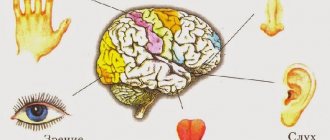
What is usually called the sense organ is the peripheral part of the analyzer. In humans, communication with the external environment is carried out using six senses: vision, hearing, taste, smell, touch and balance.
Signal discrimination.
To assess changes in intensity, temporal and spatial indicators of the stimulus, it is necessary to ensure a different reaction to a minimal difference
between stimuli.
This minimal difference is the threshold of discrimination.
According to the Weber-Fechner law
the sensation increases in proportion to the logarithm of the increase in the intensity of stimulation.
For spatial discrimination
For two signals, it is necessary that between the receptors they excite there is at least one unexcited receptor.
Visual analyzer.
The organ of vision is the most important of the senses, providing a person with up to 90% of information. The peripheral part of the analyzer - the organ of vision consists of the eyeball and auxiliary organs: eyelids, eyelashes, lacrimal glands, extraocular muscles (Fig. 235).
Rice. 235. Diagram of the structure of the eyeball: 1 - cornea; 2 - sclera; 3 - choroid; 4 - retina; 5 - anterior chamber of the eye; 6 - iris; 7 - posterior chamber of the eye; 8 - ciliary muscle; 9 - ligaments of Zinn; 10 - lens; 11 - vitreous body; 12 - blind spot; 13 - optic nerve; 14 - conjunctiva.
The wall of the eyeball consists of three membranes. The outer - white membrane (sclera) in the front of the eye is transparent, this part of it is called the cornea. Underneath the tunica albuginea is the choroid, which supplies the eye. In the anterior part, the choroid passes into the iris, which has a hole in the center - the pupil. The annular and radial muscles of the iris reflexively change the diameter of the pupil, regulating the amount of light entering the eye. The color of the eyes depends on the pigment of the iris. Next to the iris is the ciliary body, a muscle with the help of which the curvature of the lens changes, accommodation is carried out, adaptation to clear vision of objects located at different distances from the eye.
Between the cornea and the iris there is a cavity filled with moisture - the anterior chamber of the eye. The cavity between the iris and the lens is called the posterior chamber of the eye.
The third shell of the eyeball is the retina (Fig. 236). It contains light-sensitive cells - visual receptors, about 130 million rods, which provide black and white vision, and about 7 million cones, which provide information about color.
The maximum number of cones is located in the retina on the optical axis of the eye, opposite the pupil, this area is called the macula. In the place where the optic nerve leaves the eyeball, there are no receptors in the retina - a blind spot. The maximum number of rods is located on the periphery of the eye. The rods contain the visual pigment rhodopsin; a small amount of light is enough to decompose it. In cones, under the influence of light, iodopsin decomposes, but more light is needed to excite the cones.
The retina consists of several layers of cells: the outer one, adjacent to the choroid, is a layer of black pigment cells. This layer absorbs light, preventing its scattering and reflection. Then there is a layer containing rods and cones, followed by three more layers of cells.
Rice. 236. Structure of the retina: 1 - pigment cells; 2 - cones; 3 - sticks; 4 - bipolar cells; 5 - ganglion cells; 6 - amacrine cells.
The vitreous fills the entire cavity of the eye and is formed by a transparent gelatinous substance. Between the vitreous body and the posterior chamber of the eye is the lens, an elastic transparent formation in the form of a biconvex lens. The lens refracts light rays and brings them into focus on the retina. With the help of the ligaments of Zinn, it is attached to the ciliary (ciliary) muscle. Light passes through the cornea, the fluid in the anterior chamber of the eye, enters the posterior chamber through the pupil, passes through the lens, the vitreous body, and reaches the retina. Light rays undergo the greatest refraction in the cornea and lens, the image on the retina is reduced and inverted.
Accommodation is carried out due to the contraction of the ciliary muscle, while the ligaments of Zinn relax and the lens, due to its natural elasticity, becomes more convex. The ciliary muscle rests when a person looks into the distance, while the ligaments of Zinn are stretched and the lens flattens (Fig. 237).
Rice. 237. Change in the curvature of the lens: the ciliary muscle is relaxed above, contracted below.
With age, the lens often loses its elasticity and becomes flatter, the image from nearby objects goes behind the retina - senile farsightedness develops.
With congenital myopia, the eyeball is elongated, the focal length is closer to the retina and the image from distant objects is not sharp; with congenital farsightedness, the eyeball is shortened and the focal length goes behind the retina (Fig. 239).
Rice. 239. Refraction in normal (1), myopic (2) and farsighted eyes and optical correction of myopia (4) and farsightedness (5).
Nerve impulses travel along the fibers of the optic nerve to the posterior parts of the occipital lobes, and axons from the left halves of the retina of both eyes are sent to the left hemisphere, and from the right halves to the right. In this case, the axons from the medial halves intersect, forming a optic chiasm (Fig. 238).
Rice. 238. Diagram of human visual pathways: 1 - optic nerve; 2 - visual chiasm; 3 - geniculate bodies; 4 - visual cortex.
When the light intensity changes, a reflex change in the diameter of the pupil occurs. The sphincter muscles and constrictors are innervated by the parasympathetic nerves, the radial muscles, the dilators of the pupil, are innervated by the sympathetic nerves, so fear and pain lead to dilation of the pupils, it is not for nothing that they say: “Fear has big eyes.”
The cones in the retina are divided into three groups, some are excited by red light, others by green, and others by blue. If some group of cones does not work, diseases develop in which a non-human person can distinguish certain colors.
With a lack of vitamin A, the pigment rhodopsin in rods is not formed, and a person has difficulty seeing in the dark - the disease is called “night blindness”. Contamination of the mucous membrane of the eyelids (conjunctiva) leads to inflammatory processes - conjunctivitis. Reading in a moving vehicle, reading while lying down, and prolonged work with a computer lead to overwork of the ciliary muscle and a decrease in visual acuity. When working with small objects it should be at least 30-35 cm, the workplace should be well lit.
Features of the hearing organ
Human hearing organs are paired. What does this mean? A person can listen with both the right and left ear at the same time. Binaural hearing gives more information about the sound and amplifies it under certain conditions.
If the source of mechanical vibrations is at the same distance from the right and left ears, the signal volume increases by 50%. This means that in case of unilateral impairment, compensation with the help of a hearing aid of even low power significantly improves the quality of life.
Perceiving with two ears is better for determining the localization of sound. Binaural hearing gives:
- surround sound sensation;
- idea of the location of the source.
This helps you avoid danger (such as an approaching car) and isolate useful sounds from all the background noise when talking to one person in a noisy room.
Read more about hearing characteristics in this article.
If you experience any hearing problems, you must urgently undergo a hearing test using professional equipment. If you seek help in time, you have a chance to fully restore your hearing.
Auditory and vestibular analyzers
The peripheral part of the auditory analyzer consists of three parts: the outer, middle and inner ear (Fig. 240). Outer ear: auricle (cartilage inside) and external auditory canal 3.5 cm long; on the border between the outer and middle - the eardrum (0.1 mm thick).
Rice. 240. Human hearing organ: 1 - auricle; 2 - external auditory canal; 3 - eardrum; 4 - middle ear cavity; 5 - hammer; 6 - anvil; 7 - stirrup; 8 - vestibular apparatus; 9 - snail; 10 - nerve; 11 - Eustachian tube.
The middle ear is represented by an airy tympanic cavity with three auditory ossicles - the malleus, the incus and the stapes. The stapes is connected to the oval window of the membranous labyrinth. The tympanic cavity is connected to the nasopharynx by the Eustachian tube, the length of which is 3.5 cm, diameter 2 mm.
The inner ear includes a bony labyrinth, divided into three parts by two membranes: the vestibular (Reisner's) and the main (basilar) membranes; inside there is a membranous labyrinth filled with endolymph.
Rice. 242. Diagram of a transverse section of the cochlea, on which the organ of Corti is visible: 1 - scala vestibule; 2 - Reisner's (vestibular) membrane; 3 - membranous labyrinth; 4 - tectorial (integumentary membrane); 5 - basilar (main) membrane; 6 - hair cells; 7 - auditory nerve; 8 - scala tympani; 9 - organ of Corti.
The upper canal starts from the oval window and is called the scala vestibule, filled with perilymph (Fig. 241). At the top of the cochlea, with the help of an opening, it passes into the lower canal - the scala tympani, which ends with the membrane of the round window. On the main membrane is the organ of Corti, represented by receptor hair cells and the integumentary membrane located above them.
Rice. 241. Organs of balance and hearing (diagram): 1 - hammer; 2 - anvil; 3 - stirrup; 4 - eardrum; 5 – oval pouch; 6 - ampoule; 7 - semicircular canals; 8 - round bag; 9 - staircase vestibule; 10 - membranous labyrinth; 11 - scala tympani; 12 — snail (twisted into a spiral); 13 - perilymph; 14 - endolymph; 15 — round window; 16 - Eustachian tube.
In the organ of Corti there are about 24,000 hair cells located in 3-4 rows, their hairs are in contact with the movable integumentary membrane located above them. When the vestibular membrane bends, pressure is transferred to the endolymph, the main membrane moves, and the receptor cells touch the integumentary membrane (Fig. 242). Excitation arises in them, which is carried along the fibers of the auditory nerve (the conductive part of the analyzer) to the auditory zone of the cerebral cortex.
As you move away from the base to the apex, the main membrane becomes wider. High-pitched sounds cause vibrations in the main membrane at the base of the cochlea, where the membrane is shorter and thinner, low sounds are perceived by receptor cells at the top of the cochlea.
The peripheral part of the hearing organ is the organ of balance, the vestibular apparatus. It perceives the position of the body and is responsible for maintaining balance. It consists of three semicircular canals associated with the oval and round sacs (Fig. 243). Their cavities are filled with endolymph, which communicates with the endolymph of the membranous labyrinth of the cochlea.
Rice. 243 Structure of the labyrinth of the temporal bone: 1 - semicircular canals; 2 — channel ampoules; 3 - macula oval sac; 4 - macula of the round sac.
The semicircular canals are located in three mutually perpendicular planes, each with an extension - the ampulla. The ampoules contain gelatinous scallops (Fig. 244) with receptor cells that are excited by accelerated or rotational movements of the endolymph.
Rice. 244. Comb (cupula) on the left and spot (macula) on the right: 1 - cupula; 2 - hairs; 3 - receptor cells; 4 - supporting cells; 5 - nerve fiber; 6 - otolith membrane; 7 - gelatin layer.
The sacs contain spots on which receptor cells are located in a gelatinous mass, and on top there are otoliths - crystals of calcium carbonate that form the otolithic membrane. Excited by gravity, information is transmitted along the vestibular nerve to the vestibular nuclei of the pons, cerebellum, and motor nuclei of the cortex.
Functions of the hearing organ
When talking about the functions of the hearing organ, physiologists describe them in accordance with their anatomical structures. So, each department has its own specific tasks:
- catches sounds and directs them further (outer ear);
- transmits sound waves (outer and middle ear);
- protects against infections, loud sounds, damage to internal parts (outer ear, eardrum);
- transforms sound energy into electrical energy (inner ear).
The functions of hearing are evolutionarily closely related to danger notification and communication in the community. In order to maintain your ability to hear for a long time, you must follow simple rules for preventing hearing loss.